Department of Functional Diagnostics
Department of functional diagnostics is a branch of medicine dealing with mainly non-invasive diagnosis of diseases of organs and body systems, and assessment of the functionality of the damaged organs, systems and whole body. The majority of functional diagnostics methods used are safe, informative, and can often be made to different types of patients, as well as children and pregnant women. Functional diagnostics to detect diseases in preclinical stage (that is, when the disease does not bother the patient). Such early detection allows for the most effective treatment and prevent the progression of the disease. Functional diagnostics methods are widely used in clinical examinations and in healthy individuals. Currently the clinic is practically the entire arsenal of techniques that have proved effective in many international multicenter studies.
The main objective of the Division is timely, high-quality performing
non-invasive instrumental studies.
The department has the following offices:
- Electrocardiography - account number 5 (ECG)
- Echocardiography-room № 15 (EchoCG)
- Ultrasound diagnosis – room № 4, №17 (UsD)
- The function of the external breathing room number 13 (ERF)
- Echoencephalography, Rheoencephalography, Electroencephalography -
room № 7 (EchoEG, REG, EEG)
- Endoscopy – room № 18, № 19 (VideoEGDS, VideoKS, Bronchoscopy)
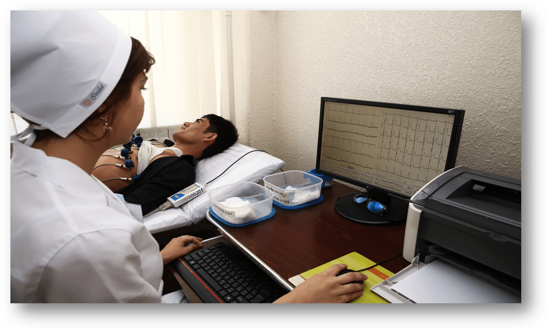
Electrocardiography
The ECG room is equipped with modern computers. Studies are conducted for ambulatory and hospital case patients. In this room Electrocardiographic studies are performed to identify coronary early signs of heart disease, myocardial infarction, congestive heart failure and arrhythmias.
The ECG room also perform rhythm gram, electrocardiogram in additional derivations.
Pharmacological tests are conducted by the instructions of attending doctors, to those patients whom physical exercises are not allowed, this test have an important role in the differential diagnosis of many cardiovascular diseases.
Echocardiography
Echocardiography is the study of the heart with the help of Ultrasound waves. During the procedure the size of the heart is examined as a whole and its individual parts, such as the ventricles, atrium and etc.
Echocardioscopy allows to examine the heart in real time, to evaluate not only its anatomy, but also contractility. Diagnostics also allows certain calculations to determine the mass of the heart, to clarify with what force and how much blood it can push through the cuts, to see different abnormalities of heart, disorder of atrium and ventricles, heart valves. If the valves are not adequately functioning or there are abnormal holes in the walls of the heart, can be seen the wrong bleeding.
Procedure Echocardioscopy is safe for anyone, even for pregnant women and infants. It does not require special training and totally painless. Now Echocardioscopy is compulsory test for all, even fully healthy children during the first year of life.
In our Clinic, Echocardioscopy is performed using latest stationary digital ultrasound high class scanner Sono Scape SSI – 6000.
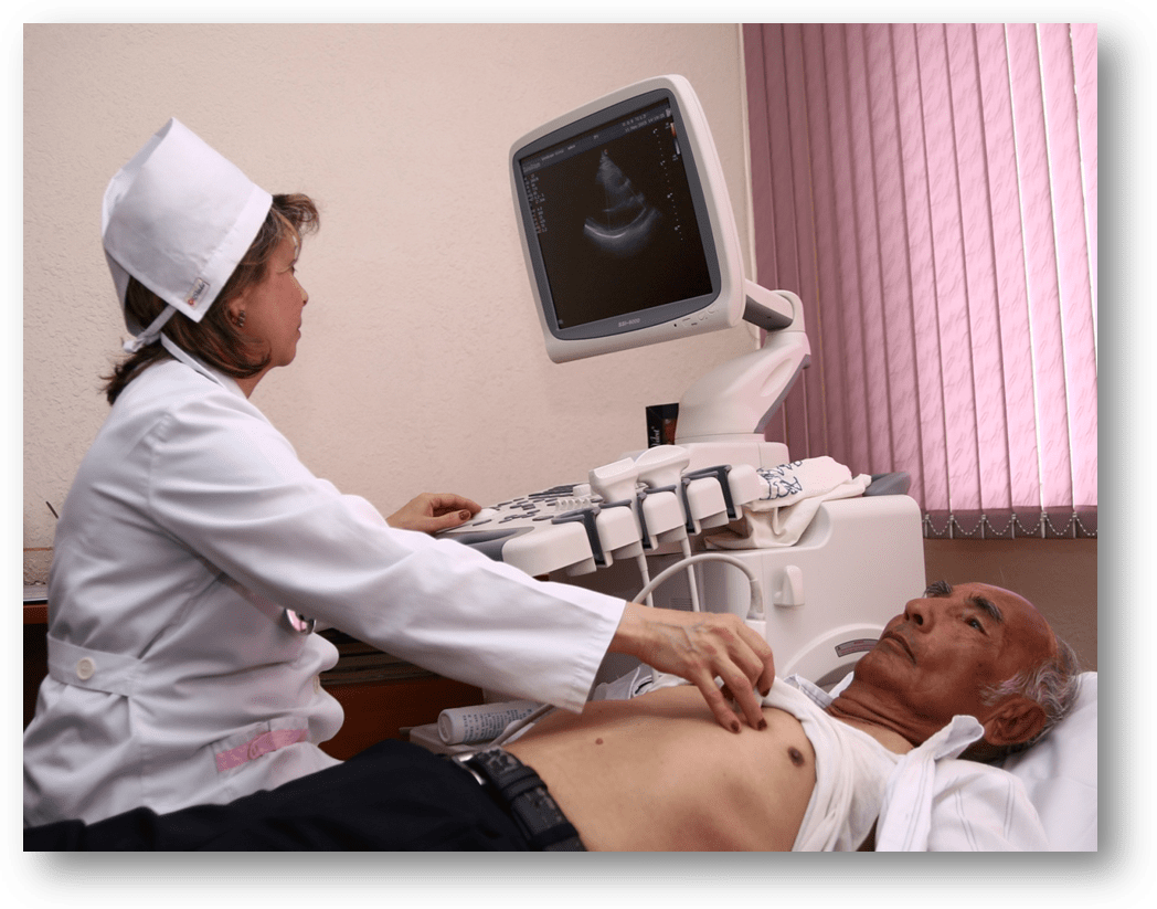
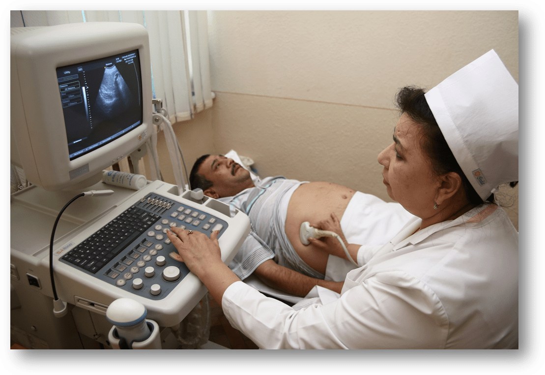
Ultrasound Diagnostics
Ultrasound diagnostics perform whole spectrum of modern ultrasound examines with help of skilled professionals using the latest generation of ultrasonic devices. All ultrasound diagnostics are conducted both ways, traditional and color Doppler mapping.
Ultrasound diagnostics are carried out on a stationary ultrasonic scanner “Sonoace 8000 LIVE” and a digital ultrasonic scanner model “DUS-6”.
Information for patients:
What research do you need? Always consult with your doctor regarding the need for an ultrasound. Remember that the final diagnosis and recommendations your doctor makes based on a variety of different data. Ultrasound help a doctor make a correct knowledge about the patient.
Preparing for diagnostics:
Some studies require special preparation: Ultrasound examination of abdominal cavity is carried out on an empty stomach-last meal should be at least 6-8 hours before the start of the study. Required condition for quality Ultrasound of the bladder and lower pelvic is when the bladder is full.
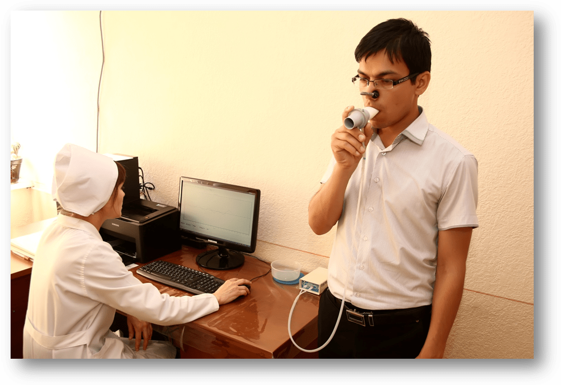
External respiratory function
Diagnostics of External Respiratory Function (ERF) are carried out on the modern computer spirograph “Spiro-Spector”, sensor which is equipped with changeable mouthpiece. It's real-time measures the speed and volume of exhaled air. Data from the sensor sanded to the computer and are processed by the program that catches the slightest deviation from the norm. Functional diagnostics physician then evaluates the source data and the product of computer analysis of spirogram, correlates with the previously completed studies and individual characteristics of the patient. The results are reflected in a detailed written opinion. Modern programs for processing ERF detect deviations from norms better than the human naked eyes. This helps us to draw conclusions, based not only on a visual assessment of ERF, but also the precise calculations in specific numbers.
A study of ERF required for the diagnosis of bronchial asthma and chronic obstructive pulmonary disease (COPD). Based on the ERF and laboratory researches we can confidently confirm or refuse the diagnosis. Evaluation of the effectiveness of treatment on changes in spirogram helps us to detect exact treatment that will have the best effect.
Preferably before the test do not eat for at least 4-5 hours, wear clothes that does not constrain breath.
Функциональние нейрофизиологические методы исследования
Functional neurophysiological methods
of Echoencephalography
Echoencephalography (study of the brain using ultrasound) is a technique for studying the anatomic relationship of the brain structures without the use of contrast agents on the basis of the principle of echolocation.
A data carrier are ultrasonic oscillations, which will help “to probe” the brain.
Echoencephalography is giving direct or indirect information on the presence of a tumor or hematoma in the brain changes occurring as a result of injury, or certain violations of brain hemodynamics, allows to record of liquor hypertension and the status of the ventricular system of the brain.
Electroencephalography
Electroencephalography (study of the functional condition of the brain)
Electroencephalography (EEG) is a method of brain diagnosis, based on registration of its electric potential that occur in the brain.
Electroencephalography - is a source of information on local and General physiological and pathological reconstructions of the functional State of the brain. For the EEG electrodes placed on the head using a number of special schemes. On the so-called system 10-20 apply 20 electrodes placed on the head in three rows.
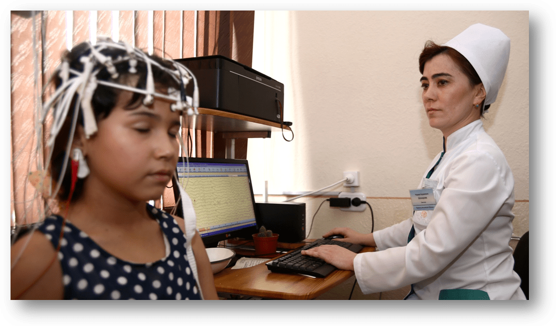
Recording bioactiveness of brain is done using standard loads (activation, photosimulation, hyperventilation).
According to testimony to the provocation epileptogenic
records using additional loads (introduction of medicinal substances).
EEG in clinical practice applied from 40 years.
Diagnostical value of the method – 80%.
Particularly valuable in epilepsy, brain injury in different rows.
Has no contraindications.
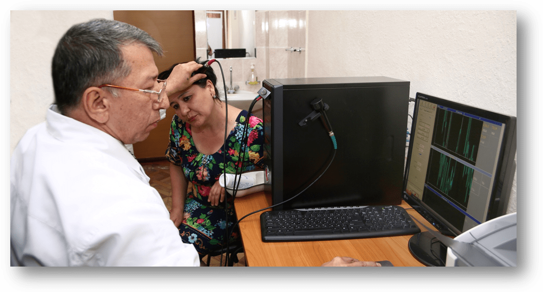
Rheoencephalography (REG)
Rheoencephalography (study of cerebral blood flow and vascular wall) method is based on the registration of electrical resistance of a brain blood formation and vascular tone. Rheoencephalography allows indirectly determine the total (outer and inner brain) blood formation, the elasticity of the arteries of the large colibri, the tone of small and medium-sized arteries of venous plethora. Method is used to estimate the passage in the carotid and vertebral arteries. Record of rheogram is standard technique and using special tests (positional, functional).
Application of standard and special samples provide a glimpse of the functional status of vascular impedance plethysmography is a valuable method for the further study of cerebral circulation. Has no contraindications, harmless.
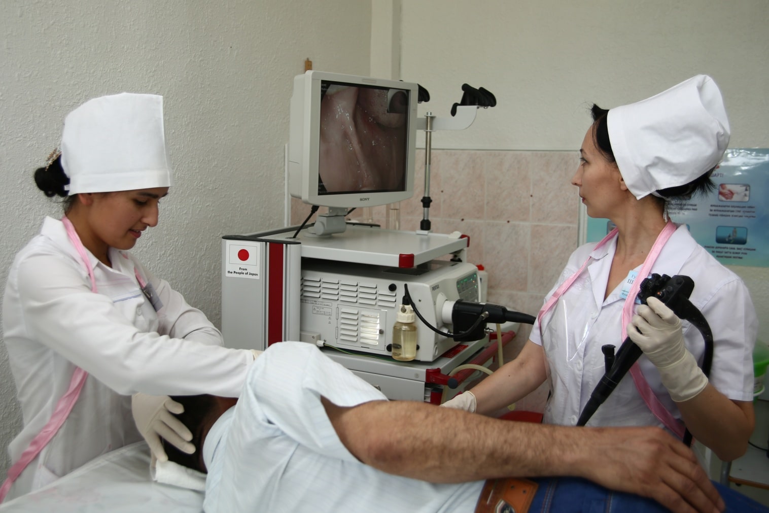
Endoscopy
Endoscopy – is visual method of study of hollow organs (through natural orifices) and cavities of the body (through punctures or operating accesses) using optical devices, equipped with lighting. If necessary, endoscopy with biopsy, x-ray or sighting ultrasound. Endoscopy is an effective and gentle method of diagnosis, one of the most efficient methods of detecting cancer of the digestive tract in the early stages of development, as well as diagnosing precancer (atrophic gastritis, stomach ulcers, polyps of the digestive tract) and treatment of endoscopically (benign tumors through an endoscope). In the Endoscopy room all kinds of diagnostic endoscopy with use of the latest digital video equipment “PENTAX” model ERC-i5000:
Video Esophagogastroduodenoscopy;
Video Colonoscopy;
Proctosigmoidoscope;
Rectosigmofibrosсopy;
Bronсhofibrosсopy.
EGDS treatment: performed by laser treatment of ulcerous disease of the esophagus, stomach and duodenal ulcers, as well as applications medicamental means (local impact).
Bronchoscopíâ ([u] bronchus (Bronchus) + Greek skopeō monitor, review; SYN. traheobronhoskopiâ) is a method of research of the inner surface of the trachea and the bronchial tubes by means of special optical devices; It is also used for a series of therapeutic actions. Diagnostic Bronchoscopy are to the pathology of the trachea, bronchi, lungs and lymph nodes intrathoracic, with blood-tinged sputum, persistent cough, shortness of breath, changes on radiographs (e.g. suspected tumor, tuberculosis, foreign body bronchi, with Pyo-inflammatory processes, bronchopulmonary system diseases of the lung and mediastinum lesion). During a bronchoscopy, in addition to exploring the mucous оболочки трахеи иthe bronchi may perform those diagnostic procedures like biopsy of the wall of the trachea, bronchus and intrabronchial (or intratracheal) located tumors; puncture biopsy peribronchial growing tumors and lymph nodes in the mediastinum; transbronchial biopsy of the pulmonary parenchyma; sampling of the contents of the respiratory tract and the bronchial tubes draining bronchial cavities for bacteriological and cytological studies; wash (lavage) small bronchi and alveoli, to study the cellular and biochemical composition of lavage fluid
Bronchoscopy helps to implement a number of measures: the bloodless removal of bronchial foreign bodies aspiration, broncholith and pathological the liquid contents of the bronchi (phlegm, blood, etc.) and thereby restore the broken road respiratory tract, elimination of asphyxia, respiratory insufficiency, etc.; refurbishment of the tracheobronchial tree in purulent bronchitis, bronchiectasias by the clearance of drugs; transbuccally drainage of abscesses of lung abscess cavity and medicines. It is an effective method of identifying the source of pulmonary bleeding and stop it.
Colonoscopy
Colonoscopy is an important method for diagnosing diseases of the large intestine
To date, colonoscopy is the most informative method of early diagnosis of benign and malignant tumors of the large intestine, nonspecific ulcerative colitis, Crohn's disease, etc. and in 80-90% of cases to examine the large intestine all over. At the time of colonoscopy visually assessed the status of the mucosa of the large intestine. When colonoscopy may also perform various therapeutic manipulation — removal of benign tumours, stopping the bleeding, removal of foreign bodies, recanalization of stenosis of intestine.
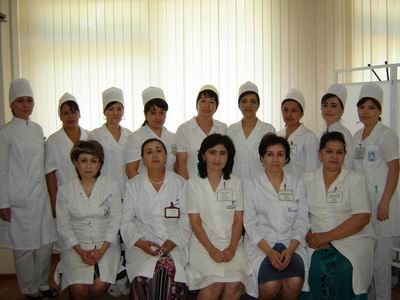
SPECIALISTS
Мирзамухамедова Умида Абраровна
Doctor USI
Олий тоифали шифокор
Қосимжонова Гулчехра Магруповна
Олий тоифали функционал диагностика шифокори
Нуритдинов Сирожиддин Сайпитдинович
Олий тоифали функционал диагностика шифокори
Шамухутдинова Гўзал Абдумаликовна
Олий тоифали эндоскопия шифокори
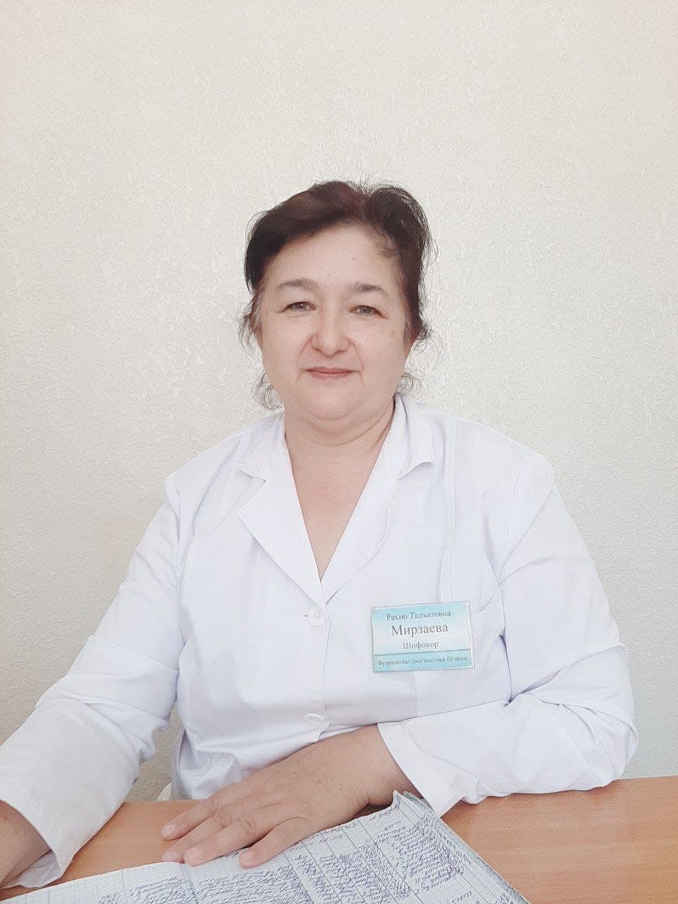
Мирзаева Раъно Таълатовна
Олий тоифали шифокор. ЭКГ

Рахимова Манзура Кутбиддиновна
Врач ЭКГ. ЭхоКГ.
Саидова Дилфуза Шоанваровна
ЭКГ врач первой категории
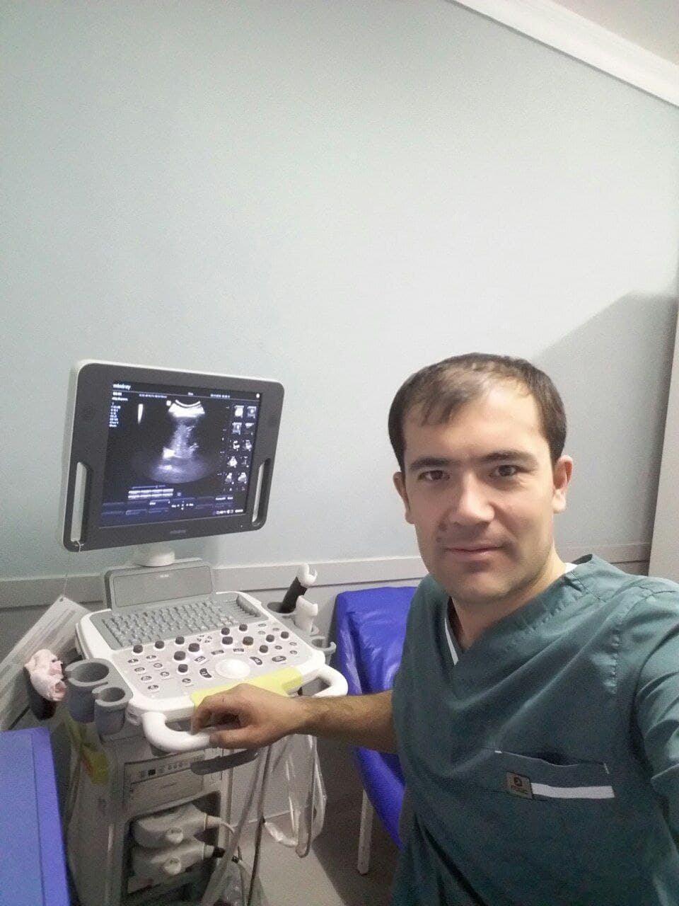
Жалолов Рустам Некмурадович
Doctor USI
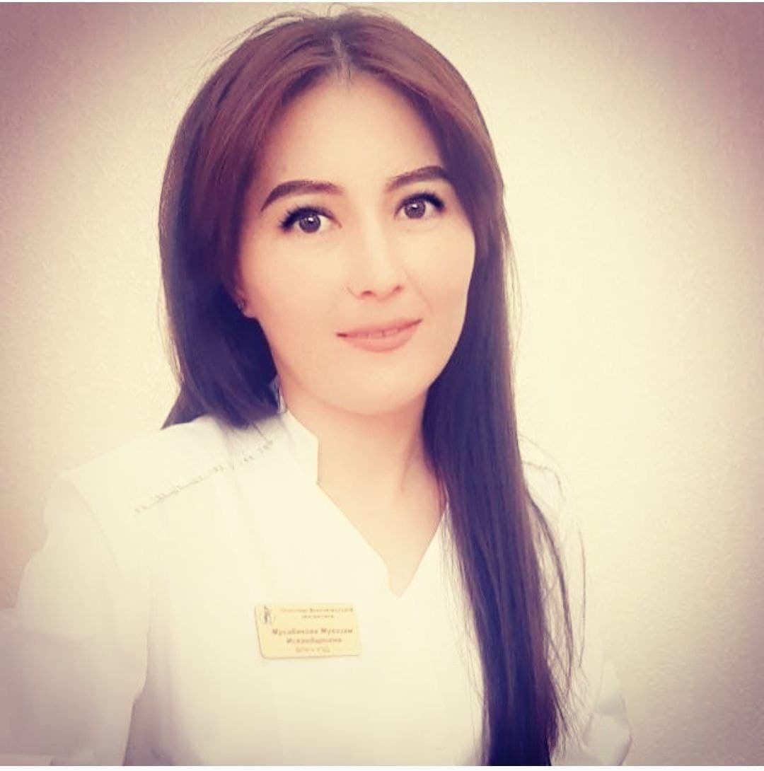
Мусабекова Муаззам Искандаровна
Doctor USI